Showing posts with label AMNH. Show all posts

Friday, July 1, 2016

Zika

The mosquito-transmitted virus Zika has been getting world-wide attention since the World Health Organization’s (WHO) announcement in February that it is a national health concern. More recently, the Zika case in New Jersey is prompting discussion about this disease in the United States. What effects does Zika have on humans? What does this mean to people around the world? What can we do to stop Zika?

An enlarged model from the American Museum of Natural History(AMNH) of Aedes aegypti,the mosquito which transmits Zika.  
Photo was taken on June 30, 2016 by author at AMNH's SciCafe event.

An average individual with no immuno-compromising disorders contracted Zika, nothing much would happen. Maybe the individual would get a headache, conjunctivitis, joint pain, or a mild fever, irritating, but not bad enough to report or go to the hospital. Most people (80%) with Zika don’t even know they have it, and completely recover after the virus leaves the body a few months later. There may be a slight connection between Guillain-Barré syndrome, which can cause severe paralysis, and Zika, but the connection is unproven and does not render Zika a national concern. Why then are people, WHO included, so worried about Zika?

Zika may go virtually unnoticed in adults, but if a fetus contacts Zika from the mother during gestation, it can develop severe birth defects such as microcephaly, increased calcification in the brain, and other motor and behavioral complications. Zika can cause birth defects no matter what trimester of pregnancy it is contracted in.
If Zika continues to spread, it could severely damage human populations. Dr. W. Ian Lipkin, John Snow Professor and Director of the Center of Infection and Immunity at Columbia University, stated at the American Museum of Natural History’s Zika SciCafe on June 30, 2016 that if there is any way to wreak havoc on a species, it is to prevent it from reproducing properly, and that’s what Zika is doing to us.

AMNH SciCafe.  Panelists at the cafe were:
 Dr. W. Ian Lipkin; Catherine Spong, MD; and Jay K. Varma MD.
Event moderator was Dr. Susan Perkins.
Photo taken at the event by author
Now, this virus with barely any noticeable symptoms sounds quite alarming, but thankfully, Zika has not yet overtaken the whole world. The virus remains restricted to southern climates such as those in South America or Africa. International travel can result in cases far from the south, however, and there have been 233 cases were reported in New York City as of July 1, 2016. Healthcare providers everywhere must be prepared to treat and prevent Zika infections. One concern about Zika being carried back to countries like the United States by travelers is that a northern relative of Aedes aegypti, the mosquito which carries Zika, will bite someone with Zika, and then continue to spread the virus to more and more people, eventually interfering with their ability to reproduce.

Where Did Zika Come From?

 Most people, myself included, only heard of Zika recently, but it has likely been around for a long time, and probably passed unnoticed until due to its generally mild symptoms. The first known case occurred in Zika forest in Uganda in the 1940s and generated little attention. In 2007, there was an outbreak on Yap Island, Micronesia which effected a high percentage of the population putting Zika on the international radar.

How Does Zika Spread?

 The main form of Zika transmission is through mosquito bites. The infection process begins when a mosquito ingests Zika-infested blood. The virus then enters the mosquito’s system, including its saliva, which transfers the virus into the next human it bites. Not all mosquitos are good transmitters of Zika. Aedes aegypti is the species of southern, aggressive, day-biting mosquito that can breed in very small amounts of stagnant water and spreads Zika. (We don’t have that species here in New York City.)

Zika can also be transmitted sexually and through blood contact. Zika remains in an infected woman’s body for about two months, and in an infected man’s semen for even longer. To prevent spreading Zika this way and causing birth defects in unborn children, couples traveling to Zika heavy-zones should be tested for Zika and avoid unprotected sex for six months, when they will both be free of the virus.

The blood contact method of transmission means Zika could be spread through blood transfusions or organ donations.

Testing for Zika

Zika is a virus, meaning it is a small packet of genetic information surrounded by proteins that can only reproduce inside of a cell. Viruses attach themselves to their host cell, penetrate the cell, unleash their genetic material, replicate it to produce new viruses, and kill the host cell.

Informational flyer from the NYC Department of Health about Zika.
Photo taken by author.
There are several ways to test for a viral infection like Zika. The preferred method is to run a polymerase chain reaction (PCR) test to determine if there is genetic evidence of the virus in the individual. The second method is to test for presence of the virus’s antibodies. The genetic test is highly accurate at determining if Zika is present, but the antibody test is much less accurate and can test positive even if another virus, not Zika, is present. The antibody test is only used because a viral infection consists of two stages: one where viral genetic information is present in the individual and one where viral antibodies are present, but not the genetic information. Between these stages is a window of time where neither genetic information nor antibodies are present. This means both viral tests are good at determining if a virus is present, but not as good at determining if it is absent. If the test comes out negative, there is still a chance that the individual could have Zika. Catherine Spong MD, Acting Director of the Eunice Kennedy Shriver National Institute of Child Health and Human Development at the National Institutes of Health and speaker at the American Museum of Natural History’s Zika SciCafe, suggests careful monitoring of pregnant women who have Zika, and those who may have contracted Zika throughout their term for the health of the baby.
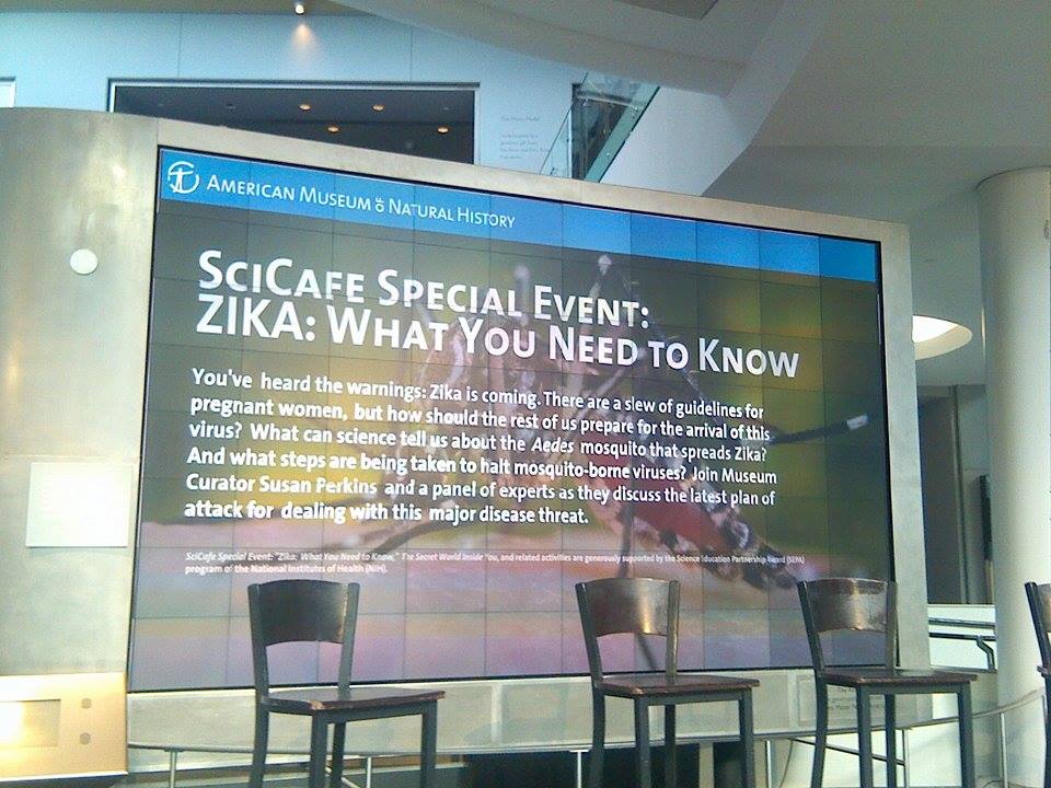
Zika SciCafe at AMNH. Photo taken by author at the event.

Prevention

 Vaccines for Zika are still being developed and tested. Even once vaccines are on the market they won’t completely eradicate the disease. There are multiple forms of Zika, and while the vaccine may successfully prevent one form, it may actually amplify the effects of a different strain. Being infected to other viral infections similar to Zika, like Dengue, can also amplify the symptoms of Zika.

Cullman Hall of the Universe at AMNH where the Zika SciCafe was held.
Photo taken by author at the event.
Another way to prevent Zika is avoid getting bitten by mosquitos. On a personal level this means wearing bug spray, long sleeved shirts and pants, putting up bug screens, and removing standing water. On a governmental scale, as Jay K. Varma MD, Deputy Commissioner for Disease Control at the New York City Department of Health and Mental Hygiene, explained at the American Museum of Natural History’s Zika SciCafe, this means activating new mosquito control programs that include putting chemicals in standing water which kill mosquito larva, spraying to kill adults, and informing the public of how to stay safe.

Here are some websites with more information on Zika:


Monday, April 25, 2016

The Importance of Images: Scientific Illustration and Photography


An etching of Caiman crocodilus and Anilius scytale by Maria Sibylla Merian
Engraver: Joseph Mulder. A public domain image.
A picture can tell the story of a thousand words. While many people may balk at reading a dense scientific article about Caiman crocodilus or Anilius scytale most of them would be happy to look at images of these animals. Images are one of the best ways to make science accessible and understandable for both laypeople and scientists.

Today illustrations and photographs are used to supplement scientific texts, but since photography didn’t exist, or was inaccessible for a large chunk of scientific history, only illustrations were used.

Woodcut print by Thomas Bewick from A History of British Birds (1797)
A public domain image
The European[1] invention of the printing press during the middle of the 15th century, as well as the advent of perspective drawing and increasing popularity of realism made the first scientific illustrations possible. The earliest illustrations were hand drawn into printed books, but as soon as illustrations could be printed and mass produced those were used instead. Often multiple people would collaborate to create an illustration, the scientist/author, the illustrator, and the printmaker. The most popular methods of printing scientific images were woodcut printing, engraving, and etching.

Copper engraving from Metamorphosis insectorum Surinamensium by  Maria Sibylla Merian 
Engraver: Joseph Mulder. A public domain image.
Umbrella Squid. Artist: unknown.
A public domain image.
American Pennyroyal woodcut print
artist: unknown. A public domain image
Most modern illustrators don’t use these printing methods anymore, but instead rely on sketches and digital touch-up methods.
When photography emerged in the 1800s it was quickly utilized by scientists. Even the earliest forms of photography, like cyanotypes and daguerreotypes were used to photograph algae and solar eclipses for scientific purposes. Science and photography have always been closely intertwined since photography is practically a science itself. Photography, especially modern digital photography, does have some benefits over illustration.
Cyanotype photograph of algae by Anna Atkins
A public domain image.
  • Photography is more accessible to the average person, practically anyone can use a camera at an amateur level, but it takes much more work to become an amateur illustrator.   
  • It takes less than a second to take a photograph, which makes it easier to accurately capture the images in the field.
  • Videos can be used to show movement that illustration can’t capture as well. 
  • Photographs can capture images not visible to the human eye. Rosalind Franklin took pictures using technology that captured invisible X-ray beams, which allowed the shape of DNA to be determined.  
  • Scientists can use motion sensor cameras placed in a study area to observe animals, and to determine what species are present in a certain area without long hours of in-person research, something illustration could never do.
  • Photography captures images as they exist in real life, the artist’s possible error in subject interpretation is not an issue.
Public domain photo provided by NASA/NSSDC

      Still, scientific illustration isn’t dead, there are still some things a drawn image can do that photography can’t.            
                                                                                               
Saturn 1874. Artist: Étienne Léopold Trouvelot.
A public domain image.

Disarticulation of the Four Fingers. Artist: Jean-Baptiste Léveillé.
A public domain image.

  • Illustrators can simplify their subject matter or emphasize important details to make complex subjects like anatomy easier to understand.
  • They can make objects transparent to do things like better show the placement of organs in the body.  
  • Illustrators can draw study skins from museum collections in life-like positions, which is great when dealing with endangered or extinct species which are near to impossible to photograph in nature.
  • Illustrators can use data from fossils to create images of ancient animals and plants as if they were still living.Illustrations show a certain sensitivity to the subject matter, lushness of color and simple layout that few photographs manage to capture. They are the ideal mixture of art and science.
Thunder and Lightning: Weather Past, Present, Future by Lauren Redniss
Author's own photo.
Artistic illustrations for popular science books, like those in Lauren Redniss’ books Thunder and Lightning: Weather Past, Present, Future and Radioactive: Marie and Pierre Curie: A Tale of Love and Fallout capture the drama and feeling of a storm and the challenge and excitement of the Curie’s lives in a way photography or even more literally accurate drawings could not.

My etching of the Opah.
About a month ago I made my own scientific illustration as part of a workshop at the American Museum of Natural History. I choose to draw and etch the Opah (Lampris guttatus, a gigantic and warm-blooded fish). I noticed that after making the etching I could remember almost exactly how the Opah looked. Observing the Opah closely enough to draw it helped me to understand the fish on a deeper level than I would have if I just read about it. Illustrations help both the viewers and the artists understand something on a deeper level than words can express.


[1] Printing was invented in China and Korea much earlier on, in the 11th and 13th centuries respectively. Europe was late to the printing game, though they did advance printing technology and use water resistant inks.

Monday, March 7, 2016

Museum Collections

 When I look at the New York City landscape around me I notice how much of it is covered with cement. It wasn’t always this way. Even now bustling Manhattan used to be rural. It makes me wonder, how has urbanization over the past century changed our environment?

Wouldn’t it be nice if we could go back fifty or a hundred years and scientifically compare their environment to ours now?

It ends up we can do just that.

Well, not exactly. Time travel has not been invented (yet!), but scientists do have a way of preserving pieces of the past for future study. Museums and scientific institutions around the world like the Smithsonian, the American Museum of Natural History, and the Natural History Museum, London, have been collecting specimens of fish, birds, invertebrates, reptiles and amphibians, and mammals for many years. These collections document biodiversity over time and across the globe.

Having these collections is a great asset to science. Let’s say a scientist wants to know how urbanization is effecting different species. The scientist could look at specimens of many different species collected from an area that was once rural but is now urbanized and examine any changes in the specimens and populations they were collected from. Maybe this scientist decides to focus how urbanization has effected one particular species. Then s/he could look a specimens of this species from different areas, and compare populations of this species over both space and time.

Once we know how urbanization has effected a species, we can use this data to predict how further development will continue to effect it in the future. Knowing what happened in the past can help people realize what needs to change if we want to maintain a healthy environment. We can use this information to develop good environmental policies and policies for conservation.

It’s pretty cool that museum collections can be used to study environmental health, but that’s not the only way collections are used. Here are a few more ways we can use species collections:

o   To study evolution. Maybe during urbanization, one population of a particular species became isolated. Using specimens collected from the original population and the isolated one, researchers can see if the populations change enough to be considered different species or different subspecies.
o   To study genetics. DNA can be obtained from tissue samples and from the skins of specimens, as long as they weren’t preserved in formalin. Today we can obtain DNA from specimens collected when people barely even knew what DNA was! Who knows what information old collections will provide scientists in the future.
o   To study disease, contaminates and parasites and how they effect a species.
o   To identify unknown species using DNA or morphology.
o   To study the diet of a species and how it changes.

The interesting thing about collections is that they are always being used in new ways. Early collectors probably had no idea the specimens they collected would be used to study the effects of urbanization. Not too long ago no one thought we would be using DNA from specimens for science, but now that is common practice. Future scientists will probably use specimens we collect today in ways on one could dream of now.

Thanks to Neil Duncan and Nuala Caomhanach at the American Museum of Natural History for helping me research for this post.

Friday, December 18, 2015

Attending a Dissection

Whenever I think I know a lot about a subject, something happens that makes me realize how little I actually know. I had a moment like last Tuesday when I went to the American Museum of Natural History to watch William Mauck III, the ornithology-laboratory supervisor at AMNH, dissect, clean and stuff a raven before it entered the collections at the museum. Mauck normally would prepare the bird alone in the lab, but this week he did the work in front of one of AMNH’s afterschool classes and a few guests like myself.
Raven pre-dissection. Photo credit: Sandra Lewocki

Anatomy is one of my favorite subjects, and I know a little about birds, so I expected what I would see during the dissection to be familiar. Mauck skinned the raven and showed us the bird’s now skinless abdomen, and I saw what I expected - large pectoral muscles and some fat stored in the clavicular region. After we all got a good look, Mauck proceeded to cut open the abdomen to get tissue samples and see what the stomach contents were.
First cuts into the raven. Photo credit: Sandra Lewocki


As he sliced open the body, all of us were shocked by what we found.

Raven being skinned. Photo credit: Sandra Lewocki
Six eggs were inside our raven! The eggs were in varying stages of development. The most developed egg had a thin shell around it while the other five did not. Since the bird had been in the freezer for so long, the eggs were hard and we got to see them clearly, which is rare. Even Mauck, who prepares birds all the time, was surprised. He said that normally you don’t see many eggs, and if you do, they tend to pop into a gooey mess right away.

The most developed egg and its shell. Photo credit: Sandra Lewocki
I knew birds laid eggs, of course, but I never thought about how that happened. Mauck explained to us that bird genitalia is completely different than that of mammals. Both male and female birds of most species reproduce, urinate, defecate, and, if female, lay eggs through the same opening called the cloaca.

During mating season, the cloaca swells, and male birds temporarily store sperm in their cloaca. While bird courtship is notoriously long and involved, actual intercourse is short, taking about a minute. During mating, the male bird balances on top of the female, who moves her tail out of the way, allowing the male to briefly touch his sperm-saturated cloaca to hers, just long enough for sperm transfer. (Aquatic bird anatomy and intercourse is slightly different.) The sperm enter the female’s sperm-storage tubules and will then fertilize eggs as ovulation occurs. Birds may mate several times during the season to increase chance of fertilization as only1-2% of sperm that enter the female’s cloaca make it to the sperm-storage tubules.

The raven and its eggs. Photo credit: Sandra Lewocki
Female birds only have one ovary and oviduct, usually the left one, with the exception of raptors which have two. No one knows why they have only one, but it means most birds can only lay one egg per day. Once the females have sperm to fertilize their eggs, eggs develop in an assembly line. First the newly-fertilized egg is released from a follicle of the ovary to the oviduct. In the oviduct, layers of yolk are formed to provide food for the young embryo. Then the egg goes to the isthmus where the shell membranes are formed. Once that’s done, the egg moves to the uterus where the hard calcified shell is made. Colors or patterns are also added in the uterus. Finally the egg travels out the cloaca and is laid.

Egg development in the body is extremely fast, taking about 24 hours per egg. Most of the embryotic development happens outside the mother’s body, after the egg is laid. Laying eggs rather than carrying their young inside the mother’s body works well for birds since it keeps the mother from becoming too weighed down to fly.

Diagram by blog author
The raven we dissected in the AMNH classroom had one large egg with a thin shell and several eggs in earlier stages of development without shells. The raven died at a bird rehab facility near Berkeley, California of a broken wing before it could lay the eggs. The bird’s life ended, but the dissection doesn’t mark the end of the story or the end of our study - the skin and tissue samples will be kept at AMNH for researchers to learn from, both now and in the future.

Thursday, December 3, 2015

We Need Bacteria

Food allergies are on the rise. If we can eradicate smallpox, why are people becoming fatally ill when exposed to food that’s supposed to be good for them? Are we forgetting about some vital aspect of human health?

I learned from Dr. Jack Gilbert at the American Museum of Natural History’s Teen SciCafe, that yes we are neglecting an important aspect of human health.

Well, it’s not exactly human health that we’re forgetting about but the health of the millions of microbes that cover our skin and inhabit every organ of our bodies except the brain. Yes, bacteria are everywhere, and yes they are good for us.

Peanut butter toast and milk, standard snack or a potential hazard?
In our society we are taught that bacteria are bad and we must kill them with soap. While this is true about some bacteria, there are many other microbes that we need to survive.

 Dr. Jack Gilbert and his colleagues have studied differences in sterile mice (mice never exposed to any bacteria) and normal mice (teeming with bacteria). They found sterile mice’s organs wouldn’t develop properly, they had no fight or flight response to stressful situations, and these mice had severe food allergies. When these sterile mice were treated with good bacteria, they regained their fight or flight response and became tolerant of foods that once poisoned them.

Bacteria are amazing, they’ve been on earth for 3.8 billion years, far longer than we have. We evolved to live with and depend on bacteria. Each of us have our own unique microbe colony which influences many bodily functions from weight gain, to food tolerance, to mental health.

Okay, now we know there’s a link between bacteria and health, now what do we do?

Can we use our knowledge to help decrease risk of food allergies? According to Dr. Gilbert the answer is yes, but we might have to change the way the medical field functions. The nature of our modern, western, medical field is one-size-fits-all. We want easy treatments that work the same way on everyone. But, everyone has a unique microbe colony. Treatments that work for me based off my microbe colony might not work for you. Now that we can figure out people’s microbiomes and understand what they mean, it might be time to go back to personalized medicine.

Dr. Gilbert and his colleagues have worked with more personalized microbe therapy for children with food allergies by introducing butyrate-producing bacteria to their intestines. The bacteria helped the children to handle trigger foods better and reduced the severity of the children’s reactions, or even ended the reactions all together.

Bacteria aren’t the cure-all but it’s about time they get attention. At the Teen SciCafe hosted by American Museum of Natural History, microbes were put in the spotlight thanks to Dr. Gilbert’s lecture. We also got to try out a new student designed card game called Gutsy all about microbes in the gut. The card game was the perfect way to set the mood for microbe talk. Now I’m very excited about bacteria! Improving microbial health can have big impacts, such as reducing risk of food allergies, but doesn’t have to be complicated, just spending more time outdoors and eating food rich in good bacteria can improve your microbiome. (Sauerkraut anyone?)

Wednesday, October 28, 2015

Let's Talk About Dioramas

What makes a science museum come to life? Art museums have it easy, as art tends to be naturally eye-catching. How can museums make science intriguing right off the bat? I believe one answer is in dioramas depicting the natural world.

Diorama background by Francis Lee Jaques from the Whitney Bird Hall at AMNH
Dioramas combine art with nature and make science museums come to life. They tell a story and allow the visitors to experience the drama of nature in places they may never visit and see animals they may never have a chance to view in their natural habitat. Dioramas are great works of art, but their artists remain virtually unknown as the focus is on the natural world, not the artistry of the painted backdrop. I, however, find the artists to be just as interesting as the subjects of the dioramas themselves. Today I’d like to focus on a particular diorama artist, Francis Lee Jaques, who was known for his excellent skills as an artist … and for his difficult personality.

Diorama background by Francis Lee Jaques from the Whitney Bird Hall at AMNH
Francis Lee Jaques drew nature, specifically birds, ever since he was a child, but he didn’t originally peruse a career as an artist. In 1917, after he had already worked as a lumberjack, electrician, and railroad fireman, Jaques enlisted in the army in San Francisco. While in California, he visited the California Academy of Sciences and became interested in becoming a museum artist. Jaques was an ambitious man and he wasn’t about to apply to just any humble museum. To start off his career, he applied for a job at the University Museum of Minnesota. Jaques sent a letter to one of the curators to say that he thought the curator’s exhibition photos were flat and in need of retouching, and that he Jaques was the man for the job. Jaques was not hired.

Diorama background by Francis Lee Jaques from the Whitney Bird Hall at AMNH
Jaques was completely undeterred and sent off an application and some of his paintings to Frank Chapman the American Museum of Natural History in New York City. This was a daring move since Jaques had no introduction to the influential Chapman. Still, Jaques’ art was impressive enough that Chapman convinced James L. Clark, the head of exhibitions, to hire Jaques without so much as an interview. In 1924 Jaques packed up and moved to NYC for his new job.

Diorama background by Francis Lee Jaques from the Whitney Bird Hall at AMNH
Jaques proved to be a remarkable artist known for producing quality work in a short amount of time. As a child Jaques spent much of his time outdoors drawing and hunting with his father. This outdoor experience allowed him to help scientists with their field work when he accompanied them on field expeditions.

Diorama background by Francis Lee Jaques from the Whitney Bird Hall at AMNH
Jaques was far from perfect, and his main weaknesses were being rude, blunt, and grudge holding. He started fights with Chapman over his pay and vacation hours, with Clark over the design of the North American Mammal Hall, and with James Perry Wilson, another diorama artist, over practically anything. Wilson was a quiet man who lived with his mother and went to classical music concerts alone. Jaques made fun of Wilson’s odd habits and noted that Wilson used his brain, but in strange ways like figuring out where to stand on the train platform in order it get a good seat on the train. All Wilson ever said about Jaques was that his painted birds were sometimes too large, a true observation.

Musk Ox diorama background by Francis Lee Jaques from the North American Mammal Hall at AMNH
Jaques worked for AMNH for 18 years and produced many beautiful works of art, but he did not end his stay there on a good note. In 1957 the museum decided Jaques’ work was too stylized and they had another artist, Matthew Kalmenoff, to retouch parts of the Glacier Park Timberland diorama. When Jaques found out, he immediately quit his job and never set foot in AMNH again. 


Diorama background by Francis Lee Jaques from The Minnesota Museum of Mining
Jaques did work with museums other than AMNH including The National Museum of Wildlife Art, The Minnesota Museum of Mining, and The Bell Museum of Natural History. Jaques may have been a difficult person to work with, but his artwork continues to inspire visitors every day.


Diorama background by Francis Lee Jaques from The Minnesota Museum of Mining
Close up of the Musk Ox diorama background

Wednesday, September 30, 2015

Anthropology: What Makes It a Science?

Let’s say you’re asked to form an image for the word “science” in your mind. Most people, myself included, would probably picture something along the lines of a person in a lab coat analyzing DNA or something. This is quite a one-sided view of science, and many scientists never wear lab coats and their jobs don’t involve DNA.

If lab coats aren’t required to make a discipline a “science,” then what is? Webster’s Dictionary defines science as “knowledge acquired by careful observation, by deduction of laws which govern charges and conditions, and by testing these deductions by experiment.” With that definition, practically anything can be studied as a science.

I find anthropology to be one of the least “sciency” of the sciences, but after hearing Dr. Laurel Kendall talk about anthropology, I started thinking about what does make anthropology a science.

I went to Dr. Laurel Kendall’s lecture Picturing Spirits in Korea at the American Museum of Natural History just last week. In her research she asks what makes Korean Shaman’s paintings sacred, rather than just works of art. How is Dr. Kendall’s question and proposed answers scientific? How is her approach different than the countless other philosophers and theologians who have contemplated similar questions?


 One answer is that she reached her conclusions through experimental data and utilization of the scientific method, but I think there’s more to it than that.

Another answer has much to do with how the results are viewed. Theologians are insiders, they study their own religion. They would take the question “what makes an object sacred” and leave the answer within the context of what adherents to their respective religion believe. Philosophers do the opposite. They take this question about sacred art out of the realm of religion and put it into more generalized concepts, but they still keep the answer within the context of belief. Comparative religious scholars compare what makes an object sacred across various religions, but still the answer is in terms of beliefs and religion.

But, when anthropologists – scientists like Dr. Kendall - ask what makes an object sacred, they take the answer in context of culture and society. For example, Dr. Kendall not only discussed what makes a painting sacred to a shaman and her followers, but how the painting is valued by secular art collectors outside of the religion and even outside of Korea With Korean culture is becoming more modernized, native Korean religion and the sacredness of the paintings are changing. This shift makes the study of change possible, and that’s where scientific methods come in. 

Tuesday, September 22, 2015

Arachnids: The Recluse Spiders

Insects are everywhere. I try not to dwell on the idea that insects are crawling on everything all the time, but entomologists seem to love the fact that there are of multitudes of insects everywhere we go. That's probably why I'm not a entomologist. Still, insects are a vital part of life, and I am interested in learning more about them.

Mediterranean recluse. Photo by Antonio Serrano
This past week I attended The New York Entomological Society’s meeting on arachnids. Biologist James Beck came up from Louisiana to The American Museum of Natural History in NYC to talk about a specific genus of arachnids the Loxosceles, commonly known as recluse spiders. James Beck studies the ranges of two species of recluse, the native brown recluse (Loxosceles reclusa) and the invasive Mediterranean recluse (Loxosceles rufescens).

Recluses aren’t rare, but the public knows very little about these spiders except the fact that they are venomous. At the lecture I learned that people, even doctors treating spider bites, tend to think every spider that isn’t a widow is a recluse. James Beck told us that misidentification of recluses went so far in a nationwide survey conducted in 2005 that people asked to send in specimens of brown recluse didn’t send in a single one. The public did send many other spider species, and even a scorpion, but no brown recluses.

I’ll admit that I would probably not be able to tell the difference between a brown recluse and a Mediterranean recluse if I saw either one. I don’t think I would mistake a scorpion for a spider like someone in the study did, but identifying Loxosceles is not an easy feat, as James Beck explained. Brown recluse and Mediterranean recluse can be distinguished by their tibial segments (a part of the leg) and mouthparts, both of which require magnification to see. Different recluse species don’t like to live side by side, so if a Mediterranean recluse is identified, there shouldn’t be any brown recluse around.

Knowing the difference between the two types of recluse might be tricky, but identifying the general genus Loxosceles isn’t as hard. Loxosceles are terrestrial spiders that like to live in places like crowded sheds and basements.


Brown Recluse Photo by Rosa Pineda
Don’t worry about finding a recluse if you don’t want to. They are named “recluse” for a reason, and Loxosceles are timid spiders. They rarely bite unless provoked and aren’t at all interested in harming humans. Spiders get a bad rap for being venomous and aggressive, but in reality they are helpful creatures that kill pests like cockroaches. To avoid being bitten, if you see a spider on you, don’t smack it, gently put it down. Try to bring out that bug loving entomologist we all have within us and both you and the spider will come out happier.
 photo of a live brown recluse from the lecture